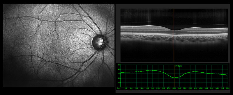
Optical Coherence Tomography (OCT)
OCT is a specialized evaluation using a laser to quantify the thickness of the optic nerve. The thicker the optic nerve, the more nerve fibers are present, and typically the healthier the optic nerve. If the fibers are progressively lost, as is the case with glaucoma, the nerve will thin and may correlate to worsening of the disease. This test is performed routinely to detect any subtle changes in the optic nerve over time.

Optic Nerve Photography
Optic nerve photographs are digital photographs taken of the optic nerve, typically when the patient’s eyes are dilated. They are performed to document how the optic nerve physically looks. The doctor will utilize these photographs for comparison on subsequent visits to check if the optic nerve is changing based on the initial picture.
Optic nerve photographs also may be used in follow-up visits to document the presence of hemorrhages on the optic nerve, which can intermittently occur over time. These pinpoint hemorrhages may be a sign of glaucoma progression. These hemorrhages resolve over time on their own and often signal to your eye doctor to adjust your glaucoma treatment.
